Le miracle africain est un sophisme. Si l’Afrique s’en sort mieux que les autres continents ce n’est pas du fait, comme certains le prétendent, que l’africain est presque un cousin lointain mais intime des virus avec lesquels il rythme son existence au quotidien. Ce n’est non plus grâce au climat africain. Car les températures observées sur notre continent existent tout aussi dans les autres parties du monde sans que cela n’ait une incidence négative réelle sur le taux de prévalence du coronavirus dans ces contrées lointaines. Même si les données démographiques servent principalement d’arguments pour tenter d’expliquer la raison pour laquelle l’hécatombe africaine annoncée n’a pas eu lieu, du moins pour le moment, je pense qu’il y a tout autre chose à l’œuvre qui n’est ni fortuite, ni empirique, ni ex nihilo. Pour peu qu’on s’y penche sérieusement alors, on s’en rendra bien compte. On pourrait ainsi expliquer le très faible taux de létalité du coronavirus en Afrique et s’en servir comme base d’orientation d’une réflexion scientifique rigoureuse locale et historicisée plutôt que globale et universelle voire universalisante sur la prise en charge de la pandémie en Afrique.
Une telle approche pourrait nous permettre de répondre à la question de savoir si nous avons vraiment besoin d’un vaccin qui, non seulement, ne nous immunise pas ; mais aussi et surtout, ne nous dispense pas du port de masque, de la distanciation sociale, de la désinfection régulière des mains ; pas plus qu’il nous rend résistant au virus et nous empêche d’être contagieux quand bien même nous serions asymptomatiques. A quoi bon alors un vaccin qui ne procure aucune immunité, incapable donc de nous empêcher de contracter le virus, de le transmette ou d’en mourir. Bref ! Un vaccin qui ne permet pas à notre système d’habitus social de poursuivre son cours après l’ouverture de la parenthèse pandémique. La seule propriété du vaccin, nous dit-on, est de nous empêcher d’en mourir en donnant à notre système immunitaire une information calibrée qui lui permettra de combattre le coronavirus avec ses propres moyens. Mais comment meurt-on à proprement parler du coronavirus ?
Qu’on se situe en anthropozoologie, en phylogénétique, en virologie, en immunologie, en hépatologie, en neurologie, en néphrologie, en cardiologie ou encore en anatomopathologie, un phénomène d’hyper-inflammation de l’organisme aussi appelé orage de cytokines est, d’après les récentes recherches sur la physiopathologie du coronavirus (SARS-CoV-2), responsable des cas sévères et partant, de la mortalité (voir références ci-dessous). Cette frénétique hyper-inflammation de notre système immunitaire engendre une cascade d’effets délétères dont une baisse brutale de la tension artérielle, des œdèmes pulmonaires, des détresses respiratoires aiguës ou encore une diminution de la force contractile du muscle cardiaque conduisant ainsi à la mort. En d’autres termes, dans le cas de la Covid-19, c’est notre propre système immunitaire qui nous tue en produisant plus d’anticorps qu’il ne devrait.
Si alors en Afrique les cas sévères ne font pas légion, c’est que quelque chose qui n’est pas d’ordre génétique car les africains n’ont pas tous les mêmes gènes ; qui va au-delà des considérations démographiques, celles-ci n’étant pas uniformes sur tous les terroirs africains ; qui n’est tout aussi pas, comme certains le prétendent, une immunité acquise au fil du temps à la suite des multiples épidémies récurrentes qui gangrènent le continent ; freine, voire empêche l’orage de cytokines au sein de notre organisme en agissant comme un immunosuppresseur. Cela veut donc dire, et il ne saurait en être autrement, qu’il doit y avoir, si ce n’est qu’il y a forcément une explication scientifique plausible à la résistance dont l’Afrique fait montre face à la pandémie qui, de plus en plus, tend à devenir une endémie. Ce sera l’objet de ma prochaine publication…
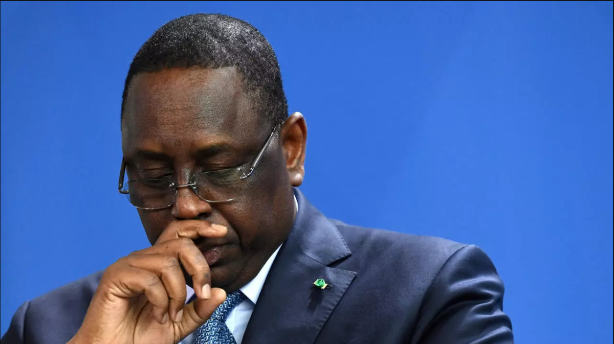
Il n’est donc pas indispensable, au-delà des capacités d’accueil limitées de nos hôpitaux, d’entraver les libertés individuelles en se couvrant du manteau de la loi ; de paralyser tous les secteurs économiques et d’hypothéquer l’avenir de nos enfants, et partant, celui du pays tout entier en plombant le système éducatif déjà fortement affecté par les décisions infructueuses, dont on sait maintenant qu’elles n’étaient pas du tout nécessaires, de fermer les écoles en mars 2020, de décaisser mille milliards dont nous ne voyons l’impact ni sur l’économie nationale, ni sur les infrastructures sanitaires et médicales. Toutes les actions entreprises depuis le début de cette crise sanitaire n’ont pas relevé d’un seul iota le niveau de vie du Sénégalais. Au contraire, tout se passe comme si le coronavirus avait atteint tous les secteurs du pays mais pas les citoyens. Couvrez-vous donc de la houppelande de la science. Elle vous conduira sereinement sur le sentier des solutions inédites, cohérentes et satisfaisantes à même de relancer notre économie tout en contenant ce que nous devons maintenant considérer comme une endémie. Bien sûr, je vous dirais pourquoi car le comment ne saurait être l’apanage de mon seul être mais le fruit d’une démarche scientifique concertée, pluridisciplinaire, transversale et holistique.
Pierre Hamet BA.
NB: Ce texte émane de recherches et d’études scientifiques que j’ai entreprises depuis le début de la pandémie SARS-CoV-2. Pour les besoins du format et pour en faciliter la lecture par un plus large public, je l’ai dépouillé de ses parties techniques et spécifiques à la médecine sans pour autant en altérer la quintessence dont les références ci-dessous constituent le fondement.
Références
1. Zhu N., Zhang D., Wang W., Li X., Yang B., Song J. A novel coronavirus from patients with pneumonia in China, 2019. N Engl J Med. 2020; 382(8):727–733.
2. Wu Y., Ho W., Huang Y., Jin D.Y., Li S., Liu S.L. SARS-CoV-2 is an appropriate name for the new coronavirus. Lancet. 2020;395(10228):949–950.
3. Wong G., Liu W., Liu Y., Zhou B., Bi Y., Gao G.F. MERS, SARS, and Ebola: the role of super-spreaders in infectious disease. Cell Host Microbe. 2015;18(4):398–401.
4. Zhou P., Yang X.L., Wang X.G., Hu B., Zhang L., Zhang W. A pneumonia outbreak associated with a new coronavirus of probable bat origin. Nature. 2020;579(7798):270–273
5. Guan W., Ni Z., Hu Y., Liang W., Ou C., He J. Clinical characteristics of coronavirus disease 2019 in China. N Engl J Med. 2020;382(18):1708–1720.
6. Huang C., Wang Y., Li X., Ren L., Zhao J., Hu Y. Clinical features of patients infected with 2019 novel coronavirus in Wuhan, China. Lancet. 2020;395:497–506.
7. Li H., Liu L., Zhang D., Xu J., Dai H., Tang N. SARS-CoV-2 and viral sepsis: observations and hypotheses. Lancet. 2020;395(10235):1517–1520.
8. Remy K.E., Brakenridge S.C., Francois B., Daix T., Deutschman C.S., Monneret G. Immunotherapies for COVID-19: lessons learned from sepsis. Lancet Respir Med. 2020:0.
9. Cui J., Li F., Shi Z.-L. Origin and evolution of pathogenic coronaviruses. Nat Rev Microbiol. 2019;17:181–192.
10. Yin Y., Wunderink R.G. MERS, SARS and other coronaviruses as causes of pneumonia. Respirology. 2018;23:130–137.
11. Ren L.-L., Wang Y.-M., Wu Z.-Q., Xiang Z.-C., Guo L., Xu T. Identification of a novel coronavirus causing severe pneumonia in human: a descriptive study. Chin Med J (Engl) 2020;133(9):1015–1024.
12. Wu F., Zhao S., Yu B., Chen Y.-M., Wang W., Song Z.-G. A new coronavirus associated with human respiratory disease in China. Nature. 2020;579:259–265.
13. De Wilde A.H., Snijder E.J., Kikkert M., van Hemert M.J. Host factors in coronavirus replication. Curr Top Microbiol Immunol. 2018;419:1–42.
14. Wrapp D., Wang N., Corbett K.S., Goldsmith J.A., Hsieh C.-L., Abiona O. Cryo-EM structure of the 2019-nCoV spike in the prefusion conformation. Science. 2020;367:1260–1263.
15. De Wit E., van Doremalen N., Falzarano D., Munster V.J. SARS and MERS: recent insights into emerging coronaviruses. Nat Rev Microbiol. 2016;14:523–534.
16. Zhang T., Wu Q., Zhang Z. Probable pangolin origin of SARS-CoV-2 associated with the COVID-19 outbreak. Curr Biol. 2020;30:1346–1351.
17. Lam T.T.-Y., Shum M.H.-H., Zhu H.-C., Tong Y.-G., Ni X.-B., Liao Y.-S. Identifying SARS-CoV-2 related coronaviruses in Malayan pangolins. Nature. 2020 DOI : 10.1038/s41586-020-2169-0.
18. Wang Q., Zhang Y., Wu L., Niu S., Song C., Zhang Z. Structural and functional basis of SARS-CoV-2 entry by using human ACE2. Cell. 2020 DOI : 10.1016/j.cell.2020.03.045.
19. Xiao K., Zhai J., Feng Y., Zhou N., Zhang X., Zou J.-J. Isolation of SARS-CoV-2-Related coronavirus from Malayan pangolins. Nature. 2020 DOI : 10.1038/s41586-020-23136-x.
20. Tang X, Wu C, Li X, Song Y, Yao X, Wu X, et al. On the origin and continuing evolution of SARS-CoV-2. Natl Sci Rev. https://doi.org/10.1093/nsr/nwaa036.
21. Van Doremalen N., Bushmaker T., Morris D.H., Holbrook M.G., Gamble A., Williamson B.N. Aerosol and surface stability of SARS-CoV-2 as compared with SARS-CoV-1. N Engl J Med. 2020;382(16):1564–1567.
22. Wölfel R., Corman V.M., Guggemos W., Seilmaier M., Zange S., Müller M.A. Virological assessment of hospitalized patients with COVID-2019. Nature. 2020 DOI : 10.1038/s41586-020-2196-x.
23. Lescure F.-X., Bouadma L., Nguyen D., Parisey M., Wicky P.-H., Behillil S. Clinical and virological data of the first cases of COVID-19 in Europe: a case series. Lancet Infect Dis. 2020 DOI : 10.1016/S1473-3099(20)30200-0.
24. Wang W., Xu Y., Gao R., Lu R., Han K., Wu G. Detection of SARS-CoV-2 in different types of clinical specimens. JAMA. 2020:e203786. DOI : 10.1001/jama.2020.3786.
25. Zheng S., Fan J., Yu F., Feng B., Lou B., Zou Q. Viral load dynamics and disease severity in patients infected with SARS-CoV-2 in Zhejiang province, China, January-March 2020: retrospective cohort study. BMJ. 2020:m1443.
26. Lamers M.M., Beumer J., van der Vaart J., Knoops K., Puschhof J., Breugem T.I. SARS-CoV-2 productively infects human gut enterocytes. Science. 2020 DOI : 10.1126/science.abc1669.
27. Dong L., Tian J., He S., Zhu C., Wang J., Liu C. Possible vertical transmission of SARS-CoV-2 from an infected mother to her newborn. JAMA. 2020:e204621. DOI : 10.1001/jama.2020.4621.
28. Hoffmann M., Kleine-Weber H., Schroeder S., Krüger N., Herrler T., Erichsen S. SARS-CoV-2 cell entry depends on ACE2 and TMPRSS2 and is blocked by a clinically proven protease inhibitor. Cell. 2020 [S0092867420302294]
29. Coutard B., Valle C., de Lamballerie X., Canard B., Seidah N.G., Decroly E. The spike glycoprotein of the new coronavirus 2019-nCoV contains a furin-like cleavage site absent in CoV of the same clade. Antiviral Res. 2020;176:104742.
30. Walls A.C., Park Y.-J., Tortorici M.A., Wall A., McGuire A.T., Veesler D. Structure, function, and antigenicity of the SARS-CoV-2 Spike glycoprotein. Cell. 2020;181(2)
31. Wang X., Xu W., Hu G., Xia S., Sun Z., Liu Z. SARS-CoV-2 infects T lymphocytes through its spike protein-mediated membrane fusion. Cell Mol Immunol. 2020:1–3.
32. Pécheur È.-I., Polyak S.J. The synthetic antiviral drug arbidol inhibits globally prevalent pathogenic viruses. Med Sci MS. 2016;32:1056–1059.
33. Khamitov R.A., Loginova S.I., Shchukina V.N., Borisevich S.V., Maksimov V.A., Shuster A.M. Antiviral activity of arbidol and its derivatives against the pathogen of severe acute respiratory syndrome in the cell cultures. Vopr Virusol. 2008;53:9–13.
34. Wang M., Cao R., Zhang L., Yang X., Liu J., Xu M. Remdesivir and chloroquine effectively inhibit the recently emerged novel coronavirus (2019-nCoV) in vitro. Cell Res. 2020;30:269–271.
35. Yao X., Ye F., Zhang M., Cui C., Huang B., Niu P. In vitro antiviral activity and projection of optimized dosing design of hydroxychloroquine for the treatment of severe acute respiratory syndrome coronavirus 2 (SARS-CoV-2) Clin Infect Dis. 2020:ciaa237. DOI: 10.1093/cid/ciaa237.
36. Al-Bari M.A.A. Chloroquine analogues in drug discovery: new directions of uses, mechanisms of actions and toxic manifestations from malaria to multifarious diseases. J Antimicrob Chemother. 2015;70:1608–1621.
37. Vincent M.J., Bergeron E., Benjannet S., Erickson B.R., Rollin P.E., Ksiazek T.G. Chloroquine is a potent inhibitor of SARS coronavirus infection and spread. Virol J. 2005;2:69.
38. Gautret P., Lagier J.-C., Parola P., Hoang V.T., Meddeb L., Mailhe M. Hydroxychloroquine and azithromycin as a treatment of COVID-19: results of an open-label non-randomized clinical trial. Int J Antimicrob Agents. 2020;49:1059.
39. Molina J.M., Delaugerre C., Le Goff J., Mela-Lima B., Ponscarme D., Goldwirt L. No evidence of rapid antiviral clearance or clinical benefit with the combination of hydroxychloroquine and azithromycin in patients with severe COVID-19 infection. Med Mal Infect. 2020 DOI : 10.1016/j.medmal.2020.03.006.
40. Geleris J., Sun Y., Platt J., Zucker J., Baldwin M., Hripcsak G. Observational study of hydroxychloroquine in hospitalized patients with Covid-19. N Engl J Med. 2020 DOI : 10.1056/NEJMoa2012410.
41. Borba M.G.S., Val F.F.A., Sampaio V.S., Alexandre M.A.A., Melo G.C., Brito M. Effect of high vs. low doses of chloroquine diphosphate as adjunctive therapy for patients hospitalized with Severe Acute Respiratory Syndrome coronavirus 2 (SARS-CoV-2) infection: a randomized clinical trial. JAMA Netw Open. 2020;3:e208857.
42. De Wilde A.H., Jochmans D., Posthuma C.C., Zevenhoven-Dobbe J.C., van Nieuwkoop S., Bestebroer T.M. Screening of an FDA-approved compound library identifies four small-molecule inhibitors of Middle East Respiratory Syndrome Coronavirus Replication in cell culture. Antimicrob Agents Chemother. 2014;58:4875–4884.
43. Changeux J-P, Amoura Z, Rey FA, Miyara M. A nicotinic hypothesis for Covid-19 with preventive and therapeutic implications. https://doi.org/10.32388/FXGQSB.
44. Stockman L.J., Bellamy R., Garner P. SARS: systematic review of treatment effects. PLoS Med. 2006;3(9):e343.
45. Choy K.-T., Wong A.Y.-L., Kaewpreedee P., Sia S.F., Chen D., Hui K.P.Y. Remdesivir, lopinavir, emetine, and homoharringtonine inhibit SARS-CoV-2 replication in vitro. Antiviral Res. 2020;178:104786.
46. Cao B., Wang Y., Wen D., Liu W., Wang J., Fan G. A trial of Lopinavir–Ritonavir in adults hospitalized with severe Covid-19. N Engl J Med. 2020;382(19):1787–1799.
47. Johnson & Johnson; 2020. Lack of evidence to support use of darunavir-based treatments for SARS-CoV-2.https://www.jnj.com/lack-of-evidence-to-support-darunavir-based-hiv-treatments-for-coronavirus
48. Agostini M.L., Andres E.L., Sims A.C., Graham R.L., Sheahan T.P., Lu X. Coronavirus susceptibility to the antiviral remdesivir (gs-5734) is mediated by the viral polymerase and the proofreading exoribonuclease. mBio. 2018;9(2) e00221–18.
49. Wang Y., Zhang D., Du G., Du R., Zhao J., Jin Y. Remdesivir in adults with severe COVID-19: a randomised, double-blind, placebo-controlled, multicentre trial. Lancet. 2020:2–9. DOI : 10.1016/S0140-6736(20)31022-9.
50. Grein J., Ohmagari N., Shin D., Diaz G., Asperges E., Castagna A. Compassionate Use of remdesivir for patients with severe Covid-19. N Engl J Med. 2020 DOI : 10.1056/NEJMoa2007016.
51. Channappanavar R., Fehr A.R., Vijay R., Mack M., Zhao J., Meyerholz D.K. Dysregulated type I interferon and inflammatory monocyte-macrophage responses cause lethal pneumonia in SARS-CoV-infected mice. Cell Host Microbe. 2016;19:181–193.
52. Siddiqi H.K., Mehra M.R. COVID-19 illness in native and immunosuppressed states: a clinical-therapeutic staging proposal. J Heart Lung Transplant. 2020;39(5):405–407.
53. Hadjadj J., Yatim N., Barnabei L., Corneau A., Boussier J., Pere H. Impaired type I interferon activity and exacerbated inflammatory responses in severe Covid-19 patients. MedRxiv. 2020 DOI : 10.1101/2020.04.19.20068015.
54. Kuba K., Imai Y., Rao S., Gao H., Guo F., Guan B. A crucial role of angiotensin converting enzyme 2 (ACE2) in SARS coronavirus–induced lung injury. Nat Med. 2005;11:875–879.
55. Singh A.K., Gupta R., Misra A. Comorbidities in COVID-19: Outcomes in hypertensive cohort and controversies with renin angiotensin system blockers. Diabetes Metab Syndr Clin Res Rev. 2020;14:283–287.
56. Vaduganathan M., Vardeny O., Michel T., McMurray J.J.V., Pfeffer M.A., Solomon S.D. Renin–angiotensin–aldosterone system inhibitors in patients with Covid-19. N Engl J Med. 2020;382(17):1653–1659.
57. Liu Y., Yang Y., Zhang C., Huang F., Wang F., Yuan J. Clinical and biochemical indexes from 2019-nCoV infected patients linked to viral loads and lung injury. Sci China Life Sci. 2020;63:364–374.
58. Mehra M.R., Desai S.S., Kuy S., Henry T.D., Patel A.N. Cardiovascular disease, drug therapy, and mortality in Covid-19. N Engl J Med. 2020 DOI : 10.1056/NEJMoa2007621.
59. Reynolds H.R., Adhikari S., Pulgarin C., Troxel A.B., Iturrate E., Johnson S.B. Renin–angiotensin–aldosterone system inhibitors and risk of Covid-19. N Engl J Med. 2020 DOI : 10.1056/NEJMoa2008975.
60. Mancia G., Rea F., Ludergnani M., Apolone G., Corrao G. Renin–angiotensin–aldosterone system blockers and the risk of Covid-19. N Engl J Med. 2020 DOI : 10.1056/NEJMoa2006923.
61. Allison S.J. SARS-CoV-2 infection of kidney organoids prevented with soluble human ACE2. Nat Rev Nephrol. 2020;1 DOI : 10.1038/s41581-020-0291-8.
62. Finlay B.B., McFadden G. Anti-immunology: evasion of the host immune system by bacterial and viral pathogens. Cell. 2006;124:767–782.
63. Vabret N., Britton G.J., Gruber C., Hegde S., Kim J., Kuksin M. Immunology of COVID-19: current state of the science. Immunity. 2020 DOI : 10.1016/j.immuni.2020.05.002.
64. Stetson D.B., Medzhitov R. Type I interferons in host defense. Immunity. 2006;25:373–381.
65. Commins S.P., Borish L., Steinke J.W. Immunologic messenger molecules: cytokines, interferons, and chemokines. J Allergy Clin Immunol. 2010;125:S53–S72.
66. Versteeg G.A., Bredenbeek P.J., van den Worm S.H.E., Spaan W.J.M. Group 2 coronaviruses prevent immediate early interferon induction by protection of viral RNA from host cell recognition. Virology. 2007;361:18–26.
67. Snijder E.J., van der Meer Y., Zevenhoven-Dobbe J., Onderwater J.J.M., van der Meulen J., Koerten H.K. Ultrastructure and origin of membrane vesicles associated with the severe acute respiratory syndrome coronavirus replication complex. J Virol. 2006;80:5927–5940.
68. Hu Y., Li W., Gao T., Cui Y., Jin Y., Li P. The severe acute respiratory syndrome coronavirus nucleocapsid inhibits type I interferon production by interfering with TRIM25-mediated RIG-I ubiquitination. J Virol. 2017:91.
69. Züst R., Cervantes-Barragan L., Habjan M., Maier R., Neuman B.W., Ziebuhr J. Ribose 2′-O-methylation provides a molecular signature for the distinction of self and non-self mRNA dependent on the RNA sensor Mda5. Nat Immunol. 2011;12:137–143.
70. Barber G.N., STING: infection, inflammation and cancer. Nat Rev Immunol. 2015;15:760–770.
71. Mesev E.V., LeDesma R.A., Ploss A. Decoding type I and III interferon signalling during viral infection. Nat Microbiol. 2019;4:914–924.
72. Devaraj S.G., Wang N., Chen Z., Chen Z., Tseng M., Barretto N. Regulation of IRF-3-dependent innate immunity by the papain-like protease domain of the severe acute respiratory syndrome coronavirus. J Biol Chem. 2007;282:32208–32221.
73. Yang Y., Zhang L., Geng H., Deng Y., Huang B., Guo Y. The structural and accessory proteins M, ORF 4a. ORF 4b, and ORF 5 of Middle East respiratory syndrome coronavirus (MERS-CoV) are potent interferon antagonists. Protein Cell. 2013;4:951–961.
74. Minakshi R., Padhan K., Rani M., Khan N., Ahmad F., Jameel S., The S.A.R.S. Coronavirus 3a protein causes endoplasmic reticulum stress and induces ligand-independent downregulation of the type 1 interferon receptor. PloS One. 2009;4:e8342.
75. Wathelet M.G., Orr M., Frieman M.B., Baric R.S. Severe acute respiratory syndrome coronavirus evades antiviral signaling: role of nsp1 and rational design of an attenuated strain. J Virol. 2007;81:11620–11633.
76. Frieman M., Ratia K., Johnston R.E., Mesecar A.D., Baric R.S. Severe acute respiratory syndrome coronavirus papain-like protease ubiquitin-like domain and catalytic domain regulate antagonism of IRF3 and NF-kappaB signaling. J Virol. 2009;83:6689–6705.
77. Menachery V.D., Gralinski L.E., Mitchell H.D., Dinnon K.H., Leist S.R., Yount B.L. Middle East respiratory syndrome coronavirus nonstructural protein 16 is necessary for interferon resistance and viral pathogenesis. mSphere. 2017;2(6):e00317–e00346.
78. Canton J., Fehr A.R., Fernandez-Delgado R., Gutierrez-Alvarez F.J., Sanchez-Aparicio M.T., García-Sastre A. MERS-CoV 4b protein interferes with the NF-κB-dependent innate immune response during infection. PLoS Pathog. 2018;14:e1006838.
79. Angeletti S., Benvenuto D., Bianchi M., Giovanetti M., Pascarella S., Ciccozzi M. COVID-2019: the role of the nsp2 and nsp3 in its pathogenesis. J Med Virol. 2020
80. Wang C., Liu Z., Chen Z., Huang X., Xu M., He T. The establishment of reference sequence for SARS-CoV-2 and variation analysis. J Med Virol. 2020.
81. Gordon D.E., Jang G.M., Bouhaddou M., Xu J., Obernier K., White K.M. A SARS-CoV- 2 protein interaction map reveals targets for drug repurposing. Nature. 2020 DOI : 10.1038/s41586-020-2286-9.
82. Zhou P., Tachedjian M., Wynne J.W., Boyd V., Cui J., Smith I. Contraction of the type I IFN locus and unusual constitutive expression of IFN- in bats. Proc Natl Acad Sci USA. 2016;113:2696–2701.
83. Wang D., Hu B., Hu C., Zhu F., Liu X., Zhang J. Clinical characteristics of 138 hospitalized patients with 2019 novel coronavirus–infected pneumonia in Wuhan, China. JAMA. 2020;323:1061–1069.
84. Blanco-Melo D, Nilsson-Payant BE, Liu W-C, Uhl S, Møller R, Jordan TX, et al. Imbalanced host response to SARS-CoV-2 drives development of COVID-19. Cell. 2020;S0092-8674(20) doi:10.1016/j.cell.2020.04.026
85. Zhou Z, Ren L, Zhang L, Zhong J, Xiao Y, Jia Z, et al. Overly Exuberant Innate Immune Response to SARS-CoV-2 Infection.(2020). Overly Exuberant Innate Immune Response to SARS-CoV-2 Infection. SSRN Electronic Journal. 10.2139/ssrn.3551623
86. Hadjadj J, Nader Yatim, Barnabei L, Corneau A, Boussier J, Pere H, et al. Impaired type I interferon activity and exacerbated inflammatory responses in severe Covid-19 patients https://www.medrxiv.org/content/10.1101/2020.04.19.20068015v1. Posted April 23, 2020 and Accessed April 27, 2020
87. Zhou F., Yu T., Du R., Fan G., Liu Y., Liu Z. Clinical course and risk factors for mortality of adult inpatients with COVID-19 in Wuhan, China: a retrospective cohort study. Lancet. 2020 [S0140673620305663]
88. Wang W., Ye L., Ye L., Li B., Gao B., Zeng Y. Up-regulation of IL-6 and TNF-alpha induced by SARS-coronavirus spike protein in murine macrophages via NF-kappaB pathway. Virus Res. 2007;128:1–8.
89. Haga S., Yamamoto N., Nakai-Murakami C., Osawa Y., Tokunaga K., Sata T. Modulation of TNF-alpha-converting enzyme by the spike protein of SARS-CoV and ACE2 induces TNF-alpha production and facilitates viral entry. Proc Natl Acad Sci USA. 2008;105:7809–7814.
90. Mehta P., McAuley D.F., Brown M., Sanchez E., Tattersall R.S., Manson J.J. COVID-19: consider cytokine storm syndromes and immunosuppression. Lancet. 2020;395:1033–1034.
91. Boudewijns R., Thibaut H.J., Kaptein S.J.F., Li R., Vergote V., Seldeslachts L. STAT2 signaling as double-edged sword restricting viral dissemination but driving severe pneumonia in SARS-CoV-2 infected hamsters. BioRxiv. 2020 DOI : 10.1101/2020.04.23.056838.
92. Ziegler C, Allon SJ, Nyquist SK, Mbano I, Miao VN, Cao Y, et al. SARS-CoV-2 Receptor ACE2 is an interferon-stimulated gene in human airway epithelial cells and is enriched in specific cell subsets across tissues.Cell. 2020;S0092-8674(20)30500-6. doi:10.1016/j.cell.2020.04.035
93. Qin C., Zhou L., Hu Z., Zhang S., Yang S., Tao Y. Dysregulation of immune response in patients with COVID-19 in Wuhan, China. Clin Infect Dis. 2020 DOI : 10.1093/cid/ciaa248.
94. Zheng M., Gao Y., Wang G., Song G., Liu S., Sun D. Functional exhaustion of Antiviral lymphocytes in COVID-19 patients. Cell Mol Immunol. 2020;17(5):533–535.
95. Zhao J., Yuan Q., Wang H., Liu W., Liao X., Su Y. Antibody responses to SARS-CoV-2 in patients of novel coronavirus disease 2019. Clin Infect Dis. 2020 DOI : 10.1093/cid/ciaa344.
96. To K.K.-W., Tsang O.T.-Y., Leung W.-S., Tam A.R., Wu T.-C., Lung D.C. Temporal profiles of viral load in posterior oropharyngeal saliva samples and serum antibody responses during infection by SARS-CoV-2: an observational cohort study. Lancet Infect Dis. 2020;20(5):565–574.
97. Guo L., Ren L., Yang S., Xiao M., Chang D., Yang F. Profiling early humoral response to diagnose novel coronavirus disease (COVID-19) Clin Infect Dis. 2020 DOI : 10.1093/cid/ciaa310.
98. Grzelak L., Temmam S., Planchais C., Demeret C., Huon C., Guivel F. SARS-CoV-2 serological analysis of COVID-19 hospitalized patients, pauci-symptomatic individuals and blood donors. medRxiv. 2020 DOI : 10.1101/2020.04.21.20068858.
99. Shen C., Wang Z., Zhao F., Yang Y., Li J., Yuan J. Treatment of 5 critically ill patients with COVID-19 with convalescent plasma. JAMA. 2020;323(16):1582–1589.
100. Luo F., Liao F.-L., Wang H., Tang H.-B., Yang Z.-Q., Hou W. Evaluation of antibody-dependent enhancement of SARS-CoV infection in rhesus macaques immunized with an inactivated SARS-CoV vaccine. Virol Sin. 2018;33:201–204.
101. Ju B., Zhang Q., Ge X., Wang R., Yu J., Shna S. Potent human neutralizing antibodies elicited by SARS-CoV-2 infection. bioRxiv. 2020 DOI : 10.1101/2020.03.21.990770.
102. Sacre K., Criswell L.A., McCune J.M. Hydroxychloroquine is associated with impaired interferon-alpha and tumor necrosis factor-alpha production by plasmacytoid dendritic cells in systemic lupus erythematosus. Arthritis Res Ther. 2012;14:R155.
103. Namiuchi S., Kumagai S., Imura H., Suginoshita T., Hattori T., Hirata F. Quinacrine inhibits the primary but not secondary proliferative response of human cytotoxic T cells to allogeneic non-T cell antigens. J Immunol. 1984;132(3):1456–1461.
104. Goldman F.D., Gilman A.L., Hollenback C., Kato R.M., Premack B.A., Rawlings D.J. Hydroxychloroquine inhibits calcium signals in T cells: a new mechanism to explain its immunomodulatory properties. Blood. 2000;95:3460–3466.
105. Picot S., Peyron F., Vuillez J.P., Polack B., Ambroise-Thomas P. Chloroquine inhibits tumor necrosis factor production by human macrophages in vitro. J Infect Dis. 1991;164:830.
106. Fordham J.N., Kirwan J., Cason J., Currey H.L. Prolonged reduction in Polymorphonuclear adhesion following oral colchicine. Ann Rheum Dis. 1981;40:605–608.
107. Chae J.J., Wood G., Richard K., Jaffe H., Colburn N.T., Masters S.L. The familial Mediterranean fever protein, pyrin, is cleaved by caspase-1 and activates NF-kappaB through its N-terminal fragment. Blood. 2008;112:1794–1803.
108. Ni Y.-N., Chen G., Sun J., Liang B.-M, Liang Z.-A. The effect of corticosteroids on mortality of patients with influenza pneumonia: a systematic review and meta-analysis. Crit Care. 2019;23(1):99.
109. Guilpain P., Chanseaud Y., Tamby M.C., Larroche C., Guillevin L., Kaveri S.V. Effets immunomodulateurs des immunoglobulines intraveineuses. Presse Med. 2004;33(17):1183–1194.
110. Richardson P., Griffin I., Tucker C., Smith D., Oechsle O., Phelan A. Baricitinib as potential treatment for 2019-nCoV acute respiratory disease. Lancet. 2020;395:e30–e31.
111. Kuzmich N.N., Sivak K.V., Chubarev V.N., Porozov Y.B., Savateeva-Lyubimova T.N., Peri F. TLR4 signaling pathway modulators as potential therapeutics in inflammation and sepsis. Vaccines. 2017;5(4):34.
112. Du L., He Y., Zhou Y., Liu S., Zheng B.-J., Jiang S. The spike protein of SARS-CoV–a target for vaccine and therapeutic development. Nat Rev Microbiol. 2009;7:226–236.
113. Wang Q., Wong G., Lu G., Yan J., Gao G.F. MERS-CoV spike protein: targets for vaccines and therapeutics. Antiviral Res. 2016;133:165–177.
114. Wang N., Shang J., Jiang S., Du L. Subunit vaccines against emerging pathogenic human coronaviruses. Front Microbiol. 2020;11:298.
115. Wu L.-P., Wang N.-C., Chang Y.-H., Tian X.-Y., Na D.-Y., Zhang L.-Y. Duration of antibody responses after severe acute respiratory syndrome. Emerg Infect Dis. 2007;13:1562.
116. Channappanavar R., Fett C., Zhao J., Meyerholz D.K., Perlman S. Virus-specific memory CD8 T cells provide substantial protection from lethal severe acute respiratory syndrome coronavirus infection. J Virol. 2014;88:11034–11044.
117. Tian X., Li C., Huang A., Xia S., Lu S., Shi Z. Potent binding of 2019 novel coronavirus spike protein by a SARS coronavirus-specific human monoclonal antibody. Emerg Microbes Infect. 2020;9:382–385.
118. Chen N., Zhou M., Dong X., Qu J., Gong F., Han Y. Epidemiological and clinical characteristics of 99 cases of 2019 novel coronavirus pneumonia in Wuhan, China: a descriptive study. Lancet. 2020;395:507–513.
119. Yin S., Huang M., Li D., Tang N. Difference of coagulation features between severe pneumonia induced by SARS-CoV2 and non-SARS-CoV2. J Thromb Thrombolysis. 2020:1–4. DOI : 10.1007/s11239-020-02105-8.
120. Tang N., Li D., Wang X., Sun Z. Abnormal coagulation parameters are associated with poor prognosis in patients with novel coronavirus pneumonia. J Thromb Haemost. 2020;18(4):844–847.
121. Helms J., Tacquard C., Severac F., Leonard-Lorant I., Ohana M., Merdji H. High risk of thrombosis in patients in severe SARS-CoV-2 infection: a multicenter prospective cohort study. Intensive Care Med. 2020:21.
122. Taylor F.B., Toh C.H., Hoots W.K., Wada H., Levi M. Scientific subcommittee on Disseminated Intravascular Coagulation (DIC) of the International Society on Thrombosis And Haemostasis (ISTH). Towards definition, clinical and laboratory criteria, and a scoring system for disseminated intravascular coagulation. Thromb Haemost. 2001;86:1327–1330.
123. Cui S., Chen S., Li X., Liu S., Wang F. Prevalence of venous thromboembolism in Patients with severe novel coronavirus pneumonia. J Thromb Haemost. 2020 DOI : 10.1111/jth.14830.
124. Middeldorp S., Coppens M., van Haaps T.F., Foppen M., Vlaar A.P., Müller M.C.A. Incidence of venous thromboembolism in hospitalized patients with COVID-19. J Thromb Haemost. 2020 DOI : 10.1111/jth.14888.
125. Lodigiani C., Iapichino G., Carenzo L., Cecconi M., Ferrazzi P., Sebastian T. Venous and arterial thromboembolic complications in COVID-19 patients admitted to an academic hospital in Milan, Italy. Thromb Res. 2020;191:9–14.
126. Klok F.A., Kruip M.J.H.A., van der Meer N.J.M., Arbous M.S., Gommers DAMPJ, Kant K.M. Incidence of thrombotic complications in critically ill ICU patients with COVID-19. Thromb Res. 2020 DOI : 10.1016/j.thromres.2020.04.013.
127. Poissy J., Goutay J., Caplan M., Parmentier E., Duburcq T., Lassalle F. Pulmonary embolism in COVID-19 patients: awareness of an increased prevalence. Circulation. 2020 DOI : 10.1161/CIRCULATIONAHA.120.047430.
128. Gando S., Levi M., Toh C.-H. Disseminated intravascular coagulation. Nat Rev Dis Primer. 2016;2:16037.
129. Ranucci M., Ballotta A., Di Dedda U., Bayshnikova E., Dei Poli M., Resta M. The procoagulant pattern of patients with COVID-19 acute respiratory distress syndrome. J Thromb Haemost. 2020 DOI : 10.1111/jth.14854.
130. Mussbacher M., Salzmann M., Brostjan C., Hoesel B., Schoergenhofer C., Datler H. Cell type-specific roles of NF-κB linking inflammation and thrombosis. Front Immunol. 2019;10:85.
131. Biemond B.J., Levi M., Ten Cate H., Van der Poll T., Büller H.R., Hack C.E. Plasminogen activator and plasminogen activator inhibitor I release during experimental endotoxaemia in chimpanzees: effect of interventions in the cytokine and coagulation cascades. Clin Sci1979. 1995;88:587–594.
132. Wu Y.P., Wei R., Liu Z.H., Chen B., Lisman T., Ren D.L. Analysis of thrombotic Factors in severe acute respiratory syndrome (SARS) patients. Thromb Haemost. 2006;96:100–101.
133. Gralinski L.E., Bankhead A., Jeng S., Menachery V.D., Proll S., Belisle S.E. Mechanisms of severe acute respiratory syndrome coronavirus-induced acute lung injury. mBio. 2013;4(4):e00271–e00273.
134. He L., Ding Y., Zhang Q., Che X., He Y., Shen H. Expression of elevated levels of pro-inflammatory cytokines in SARS-CoV-infected ACE2+ cells in SARS patients: relation to the acute lung injury and pathogenesis of SARS. J Pathol. 2006;210:288–297.
135. Idell S., Coagulation fibrinolysis, and fibrin deposition in acute lung injury. Crit Care Med. 2003;31:S213–S220.
136. Gupta N., Zhao Y.-Y., Evans C.E. The stimulation of thrombosis by hypoxia. Thromb Res. 2019;181:77–83.
137. Varga Z., Flammer A.J., Steiger P., Haberecker M., Andermatt R., Zinkernagel A.S. Endothelial cell infection and endotheliitis in COVID-19. Lancet. 2020;395:1417–1478.
138. Hamming I., Timens W., Bulthuis M.L.C., Lely A.T., Navis G.J., van Goor H. Tissue distribution of ACE2 protein, the functional receptor for SARS coronavirus. A first step in understanding SARS pathogenesis. J Pathol. 2004;203:631–637.
139. Monteil V., Kwon H., Prado P., Hagelkrüys A., Wimmer R.A., Stahl M. Inhibition of SARS-CoV-2 infections in engineered human tissues using clinical-grade soluble human ACE2. Cell. 2020 DOI : 10.1016/j.cell.2020.04.004. S0092-8674.
140. Magro C., Mulvey J.J., Berlin D., Nuovo G., Salvatore S., Harp J. Complement associated microvascular injury and thrombosis in the pathogenesis of severe COVID-19 infection: a report of five cases. Transl Res. 2020 DOI : 10.1016/j.trsl.2020.04.007. S1931-5244.
141. Luecke T., Pelosi P. Clinical review: positive end-expiratory pressure and cardiac output. Crit Care. 2005;9(6):607–621.
142. Bikdeli B., Madhavan M.V., Jimenez D., Chuich T., Dreyfus I., Driggin E. COVID-19 and thrombotic or thromboembolic disease: implications for prevention, antithrombotic therapy, and follow-up. J Am Coll Cardiol. 2020 DOI : 10.1016/j.jacc.2020.04.031. S0735-1097.
143. Vicenzi E., Canducci F., Pinna D., Mancini N., Carletti S., Lazzarin A. Coronaviridae and SARS-associated coronavirus strain HSR1. Emerg Infect Dis. 2004;10:413–418.
144. Tang N., Bai H., Chen X., Gong J., Li D., Sun Z. Anticoagulant treatment is associated with decreased mortality in severe coronavirus disease 2019 patients with coagulopathy. J Thromb Haemost. 2020;18(5):1094–1099.
145. Iba T., Levy J.H., Warkentin T.E., Thachil J., van der Poll T., Levi M. Diagnosis and management of sepsis-induced coagulopathy and disseminated intravascular coagulation. J Thromb Haemost. 2019;17:1989–1994.
146. Wang J., Hajizadeh N., Moore E.E., McIntyre R.C., Moore P.K., Veress L.A. Tissue Plasminogen Activator (tPA) treatment for COVID-19 associated Acute Respiratory Distress Syndrome (ARDS): a case series. J Thromb Haemost. 2020 DOI : 10.1111/jth.14828.
147. Rao S., Lau A., So H.-C. Exploring diseases/traits and blood proteins causally related to expression of ACE2, the putative receptor of 2019-nCov: a Mendelian randomization analysis. medRxiv. 2020 DOI : 10.1101/2020.03.04.20031237.
148. SARS-CoV-2 related proteins – The Human Protein Atlas. https://www.proteinatlas.org/humanproteome/sars-cov-2 (Accessed April 25, 2020).
149. Hikmet F., Méar L., Uhlén M., Lindskog C. The protein expression profile of ACE2 in human tissues. bioRwiv. 2020 DOI : 10.1101/2020.03.31.
150. Plaçais L., Richier Q. COVID-19: caractéristiques cliniques, biologiques et radiologiques chez l’adulte, la femme enceinte et l’enfant. Une mise au point au cœur de la pandémie. Rev Med Interne. 2020; 41(5):308–318.
151. Wichmann D., Sperhake J.P., Lütgehetmann M., Steurer S., Edler C., Heinemann A. Autopsy findings and venous thromboembolism in patients with COVID-19: a prospective Cohort Study. Ann Intern Med. 2020 DOI : 10.7326/M20-2003.
152. Zhang Q., Cong M., Wang N., Li X., Zhang H., Zhang K. Association of angiotensin-converting enzyme 2 gene polymorphism and enzymatic activity with essential hypertension in different gender. Medicine (Baltimore) 2018;97(42):e1291.
153. Ciaglia E., Vecchione C., Puca A.A. COVID-19 infection and circulating ACE2 levels: protective role in women and children. Front Pediatr. 2020:8.
154. Jin Y., Yang H., Ji W., Wu W., Chen S., Zhang W. Virology, epidemiology, pathogenesis, and control of COVID-19. Viruses. 2020;12:372.
155. Yang X., Yu Y., Xu J., Shu H., Xia J., Liu H. Clinical course and outcomes of critically ill patients with SARS-CoV-2 pneumonia in Wuhan, China: a single-centered, retrospective, observational study. Lancet Respir Med. 2020 [S2213260020300795]
156. Xu Z., Shi L., Wang Y., Zhang J., Huang L., Zhang C. Pathological findings of COVID-19 associated with acute respiratory distress syndrome. Lancet Respir Med. 2020;8:420–422.
157. Fox S.E., Akmatbekov A., Harbert J.L., Li G., Brown J.Q., Vander Heide R.S. Pulmonary and cardiac pathology in Covid-19: the first autopsy series from New Orleans. medRxiv. 2020 DOI : 10.1101/2020.04.06.20050575.
158. Ling Y., Xu S.-B., Lin Y.-X., Tian D., Zhu Z.-Q., Dai F.-H. Persistence and clearance of viral RNA in 2019 novel coronavirus disease rehabilitation patients. Chin Med J. 2020;133(9):1039–1043.
159. Guo Y., Korteweg C., McNutt M.A., Gu J. Pathogenetic mechanisms of severe acute respiratory syndrome. Virus Res. 2008;133:4–12.
160. Wu C., Chen X., Cai Y., Xia J., Zhou X., Xu S. Risk factors associated with acute respiratory distress syndrome and death in patients with coronavirus disease 2019 pneumonia in Wuhan, China. JAMA Intern Med. 2020:e200994. DOI : 10.1001/jamainternmed.2020.0994.
161. Richardson S., Hirsch J.S., Narasimhan M., Crawford J.M., McGinn T., Davidson K.W. Presenting characteristics, comorbidities, and outcomes among 5700 patients hospitalized with COVID-19 in the New York City area. JAMA. 2020:e206775. DOI: 10.1001/jama.2020.6775.
162. Feng G., Zheng K.I., Yan Q.-Q., Rios R.S., Targher G., Byrne C.D. COVID-19 and liver dysfunction: current insights and emergent therapeutic strategies. J Clin Transl Hepatol. 2020;8:18–24.
163. Li Y.-C., Bai W.-Z., Hashikawa T. The neuroinvasive potential of SARS-CoV2 may play a role in the respiratory failure of COVID-19 patients. J Med Virol. 2020:10. DOI : 10.1002/jmv.25728.
164. Li Y.-C., Bai W.-Z., Hashikawa T. Response to Commentary on “The neuroinvasive potential of SARS-CoV-2 may play a role in the respiratory failure of COVID-19 patients” J Med Virol. 2020 DOI : 10.1002/jmv.25960.
165. Mizuiri S., Ohashi Y. ACE and ACE2 in kidney disease. World J Nephrol. 2015;4:74–82.
166. Arentz M., Yim E., Klaff L., Lokhandwala S., Riedo F.X., Chong M. Characteristics and outcomes of 21 critically ill patients with COVID-19 in Washington State. JAMA. 2020;323(16):1612–1614. DOI : 10.1001/jama.2020.4326.
167. Cheng Y., Luo R., Wang K., Zhang M., Wang Z., Dong L. Kidney disease is associated with in-hospital death of patients with COVID-19. Kidney Int. 2020;97:829–838.
168. Cao M., Zhang D., Wang Y., Lu Y., Zhu X., Li Y. Clinical features of patients infected with the 2019 novel coronavirus (COVID-19) in Shanghai, China. medRxiv. 2020 DOI : 10.1101/2020.03.04.20030395.
169. Ransick A., Lindström N.O., Liu J., Zhu Q., Guo J.-J., Alvarado G.F. Single-cell profiling reveals sex, lineage, and regional diversity in the mouse kidney. Dev Cell. 2019;51:399–413.
170. Su H., Yang M., Wan C., Yi L.-X., Tang F., Zhu H.-Y. Renal histopathological analysis of 26 postmortem findings of patients with COVID-19 in China. Kidney Int. 2020 DOI : 10.1016/j.kint.2020.04.003. [S0085-2538(20)30369-0]
171. Aghagoli G., Gallo Marin B., Soliman L.B., Sellke F.W. Cardiac involvement in COVID-19 patients: risk factors, predictors, and complications: A review. J Card Surg. 2020 [jocs.14538]
172. Inciardi R.M., Lupi L., Zaccone G., Italia L., Raffo M., Tomasoni D. Cardiac involvement in a patient with coronavirus disease 2019 (COVID-19) JAMA Cardiol. 2020 DOI : 10.1001/jamacardio.2020.1096.
173. Hua A., O’Gallagher K., Sado D., Byrne J. Life-threatening cardiac tamponade complicating myo-pericarditis in COVID-19. Eur Heart J. 2020:ehaa253.
174. Shi S., Qin M., Shen B., Cai Y., Liu T., Yang F. Association of cardiac injury with mortality in hospitalized patients with COVID-19 in Wuhan, China. JAMA Cardiol. 2020:e200950. DOI : 10.1001/jamacardio.2020.0950.
175. Yang J.K., Feng Y., Yuan M.Y., Yuan S.Y., Fu H.J., Wu B.Y. Plasma glucose levels and diabetes are independent predictors for mortality and morbidity in patients with SARS. Diabet Med. 2006;23:623–628.
176. Yang J.-K., Lin S.-S., Ji X.-J., Guo L.-M. Binding of SARS coronavirus to its receptor damages islets and causes acute diabetes. Acta Diabetol. 2010;47:193–199.
177. Chhabra K.H., Chodavarapu H., Lazartigues E. Angiotensin converting enzyme 2: a new important player in the regulation of glycemia. IUBMB Life. 2013;65:731–738.
178. Li Z., Liu G., Wang L., Liang Y., Zhou Q., Wu F. From the insight of glucose metabolism disorder: oxygen therapy and blood glucose monitoring are crucial for quarantined COVID-19 patients. Ecotoxicol Environ Saf 2020; 2020;197:110614.
179. Butler S.O., Btaiche I.F., Alaniz C. Relationship between hyperglycemia and infection in critically ill patients. Pharmacotherapy. 2005;25(7):963–976.
180. Barton L.M., Duval E.J., Stroberg E., Ghosh S., Mukhopadhyay S. COVID-19 Autopsies, Oklahoma, USA. Am J Clin Pathol. 2020;153(6):725–733.
181. Bouaziz J.D., Duong T., Jachiet M., Velter C., Lestang P., Cassius C. Vascular skin symptoms in COVID-19: a French observational study. J Eur Acad Dermatol Venereol. 2020 DOI : 10.1111/jdv.16544.
182. Navel V., Chiambaretta F., Dutheil F. Haemorrhagic conjunctivitis with pseudomembranous related to SARS-CoV-2. Am J Ophthalmol Case Rep. 2020 DOI : 10.1016/j.ajoc.2020.100735.
183. Siedlecki J., Brantl V., Schworm B., Mayer W.J., Gerhardt M., Michalakis S. COVID-19: ophthalmological Aspects of the SARS-CoV 2 Global Pandemic. Klin Monbl Augenheilkd. 2020:1164–9381. DOI : 10.1055/a-1164-9381.
184. Wu P., Duan F., Luo C., Liu Q., Qu X., Liang L. Characteristics of ocular findings of patients with coronavirus disease 2019 (COVID-19) in Hubei Province, China. JAMA Ophthalmol. 2020:e201291. DOI : 10.1001/jamaophthalmol.2020.1291.
185. Luis E. Escobar, View ORCID ProfileAlvaro Molina-Cruz, and Carolina Barillas-Mury. BCG vaccine protection from severe coronavirus disease 2019 (COVID-19). PNAS July 28, 2020 117 (30) 17720-17726; first published July 9, 2020; updated October 12, 2020 https://doi.org/10.1073/pnas.2008410117. Published By: National Academy of Sciences Print ISSN: 0027-8424 Online ISSN: 1091-6490

2 réponses sur « MACKY SALL, ÉCOUTE-MOI TRÈS BIEN ! »
Merci à mon frère PHD pour ce texte d’envergure scientifique. Espérons que ces belles lignes tombent sous les yeux de notre Président qui semble être complètement perdu avec ses décisions à l’emporte-pièce à propos de cette maladie du coronavirus qui plombe notre économie.
Santé et longue vie mon frère !
Merci Cher Monsieur Pierre Hamet Ba!
Je vous remercie pour votre commentaire qui justifie mes propres idées.
Pour moi, pas de Vaccin.
Très respectueuselent vôtre.
Mme SAJOUS Josette.